
Histological Changes in Some Organs of The Female Rats Infected With Toxoplasma Gondii Parasite Isolated From Embryo of Aborted Sheep

Article Sidebar
-
Toxoplasma gondii, histopathological changes, Sheep
Abstract
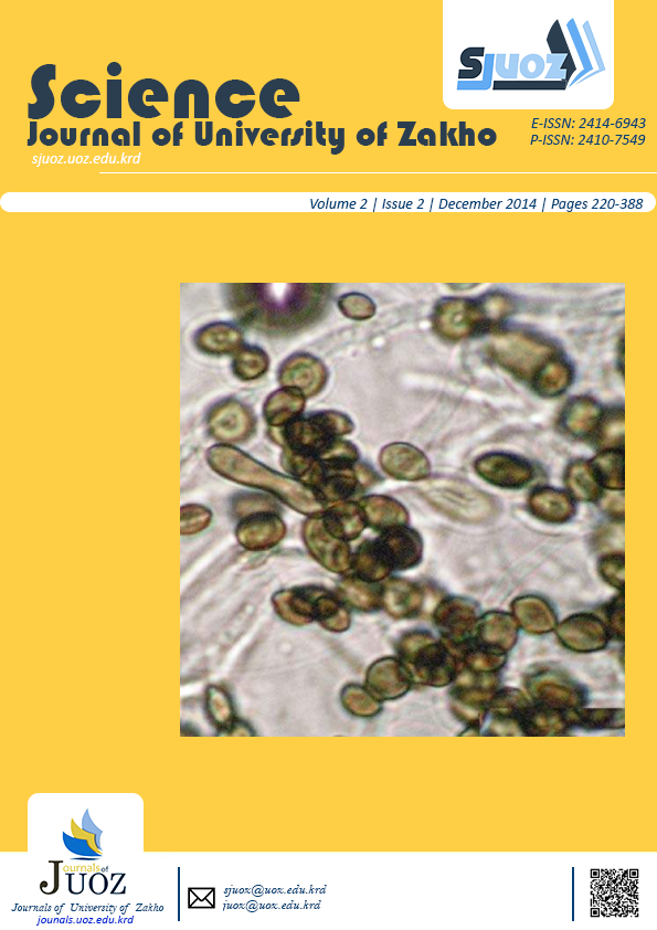
The current study included identifying the lesions and the histological changes caused by the intracellular parasite Toxoplasma gondii, which was isolated from embryos of aborted ewes. Intraperitonially injected in 15 female albino rats (3 months old) with suspension containing 100 tissue cysts.Histological section showed after four months of injection a chronic infection characterized by autolysis in all sections, where the liver sections showed expansion in the central hepatic veins, congestion in sinusoidal curves with irregularity in hepatic cords, and the presence of parasites in the liver cells. The brain tissue showed vacuoles in the neurons with an increase in the number of Purkenji cells. Kidney sections were characterized by degenerative and necrotic changes in the endothelial cells of the proximal and distal convoluted tubules with Sloughing of necrotic and degenerated cells of the tubes which accumulated inside the cavity. The parasites appeared in the endothelial cells of the glomerular tufts. Ovary and the uterus showed increased vascular wall thickness, furthermore, the spleen showed autolytic changes, deposition of pigment with the presence of parasites within the cells.
Full text article
References
Authors
Copyright (c) 2014 Liqaa H. Al-Delami, Buthaina H. Al-Sabawi, Firas M. Basheer, Hafidh I. Al-Sadi

This work is licensed under a Creative Commons Attribution 4.0 International License.
Authors who publish with this journal agree to the following terms:
- Authors retain copyright and grant the journal right of first publication with the work simultaneously licensed under a Creative Commons Attribution License [CC BY-NC-SA 4.0] that allows others to share the work with an acknowledgment of the work's authorship and initial publication in this journal.
- Authors are able to enter into separate, additional contractual arrangements for the non-exclusive distribution of the journal's published version of the work, with an acknowledgment of its initial publication in this journal.
- Authors are permitted and encouraged to post their work online.