MICROSCOPIC ARCHITECTURE OF THE RESPIRATORY AND CONDUCTING SYSTEM OF THE LUNG OF NILE MONITOR
Abstract
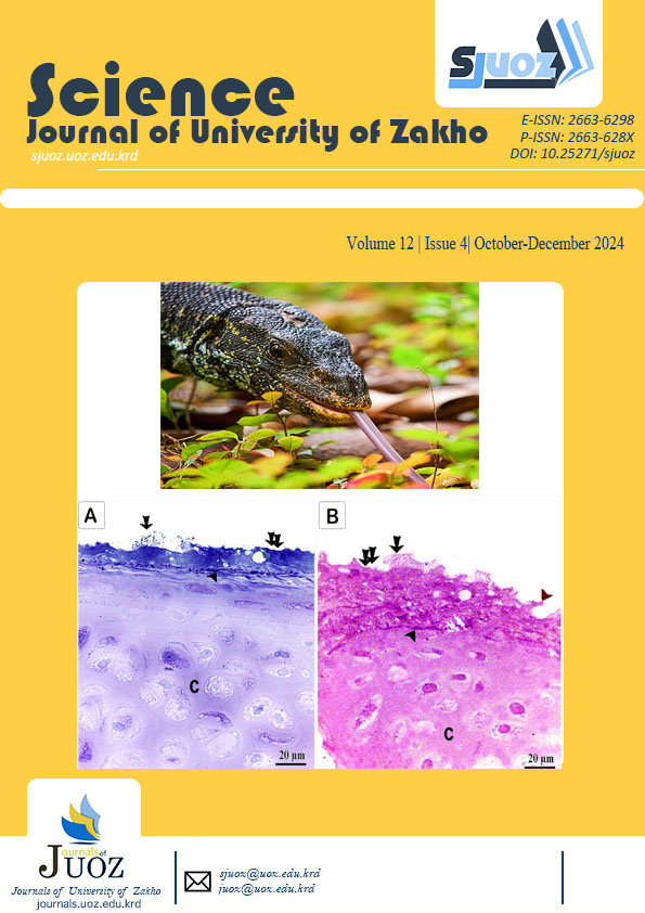
Using semi-thin sections, the present investigation examined the microscopic characteristics of the lungs of the Nile monitor (Varanus niloticus). The lungs were composed of Intrapulmonary conducting airways and respiratory faveoli. Intrapulmonary airways originate from the terminal portion of the bronchus, which extends into the lung to create the bronchial tree. The bronchus was lined with pseudostratified ciliated epithelium composed of both ciliated and non-ciliated cells, and it was supported by plates of hyaline cartilage. The central lumen is surrounded by contractile fibers that contain smooth muscle cell bundles and are covered by ciliated and non-ciliated cells. The central lumen communicates with the faveoli. Separating adjacent faveoli are pulmonary trabeculae covered with various cell types, including type I pneumocytes, type II pneumocytes, and pulmonary macrophages. Some substantial pulmonary bronchi were also supported by small cartilage plate granules and lined with ciliated epithelium. Type I pneumocytes were flat cells, whereas type II pneumocytes had cuboidal cells with vacuolated cytoplasm. Surface irregularity and vacuolated cytoplasm were features of pulmonary macrophages. In addition, the connective tissue of the pulmonary septa contained immune cells, such as Mast and Eosinophils. In conclusion, the microstructure of the lung of the Nile monitor closely resembles that of other reptile species. However, the distinction between intrapulmonary cartilage palates and pulmonary septa raises the concept of species differentiation. In addition, the discovery of various types of pulmonary immune cells enhances the Nile monitor's ability to persist in a variety of environments by enhancing its pulmonary immunity.
Full text article
References
Abd-Elhafeez, H., Moustafa, M., Zayed, A., & Sayed, R. (2017a). Morphological and morphometric study of the development of leydig cell population of donkey (Equus asinus) testis from birth to maturity. Cell Biol, 6(1), 2. DOI: 10.4172/2157-7099.1000370.
Abd-Elhafeez, H. H., & Soliman, S. A. (2017b). New description of telocyte sheaths in the bovine uterine tube: An immunohistochemical and scanning microscopic study. Cells Tissues Organs, 203(5), 295-315. DOI:10.1159/000452243.
Abd‐Elhafeez, H. H., Rutland, C. S. and Soliman, S. A. (2023). Morphology of migrating telocytes and their potential role in stem cell differentiation during cartilage development in catfish (Clarias gariepinus). Microscopy Research and Technique, 86(6),1108-1121.DOI: 10.1002/jemt.24374. Epub 2023 Jun 20.
Divers, S. J., & Mader, D. R. (2005). Reptile Medicine and Surgery - E-Book. Elsevier Health Sciences. https://books.google.com.eg/books?id=7Ai4BKhi0VUC
Dowell, S. A., Portik, D. M., de Buffrénil, V., Ineich, I., Greenbaum, E., Kolokotronis, S.-O., & Hekkala, E. R. (2016). Molecular data from contemporary and historical collections reveal a complex story of cryptic diversification in the Varanus (Polydaedalus) niloticus Species Group. Molecular Phylogenetics and Evolution, 94, 591-604. DOI: 10.1016/j.ympev.2015.10.004
Hermida, G., Farías, A., & Fiorito, L. (2003). Ultrastructural characteristics of the lung of Melanophryniscus stelzneri stelzneri (Weyenberg, 1875) (Anura, Bufonidae). Biocell : official journal of the Sociedades Latinoamericanas de Microscopía Electronica ... et. al, 26, 347-355. DOI:10.32604/biocell.2002.26.347
Abd-Elhafeez, H., Moustafa, M., A., & Sayed, R. (2017b). The development of the intratesticular excurrent duct system of donkey (Equus asinus) from birth to maturity. Histology, Cytology and Embryology, 1, 1-8. DOI: 10.15761/HCE.1000108.
South African National Biodiversity Institute. (n.d.). Nile monitor. CITES Species Database. Retrieved from https://www.sanbi.org/animal-of-the-week/nile-monitor/
Smithsonian National Zoo. (n.d.). Nile monitor. Retrieved from https://www.sanbi.org/animal-of-the-week/nile-monitor/
Lloyd, R. V. (2001). Morphology methods: cell and molecular biology techniques. Springer Science & Business Media.
Moustafa, M. A. M., Ismail, M. N.-E., Mohamed, A. E. S. A., Ali, A. O., & Tsubota, T. (2013). Hematologic and biochemical parameters of free-ranging female Nile monitors in Egypt. Journal of wildlife diseases, 49(3), 750-754. DOI: 10.7589/2013-01-002.
Nussbaum, R. (2002). A Field Guide to the Reptiles of East Africa: Kenya, Tanzania, Uganda, Rwanda and Burundi . By Stephen Spawls , Kim Howell , Robert Drewes , and James Ashe ; Consultants: Alex Duff‐MacKay and Harald Hinkel . San Diego (California): Academic Press . $49.95. 543 p; ill.; scientific and common name indexes. ISBN: 0–12–656470–1. 2002. The Quarterly Review of Biology, 77, 339-340. DOI:10.1086/345220
Peixoto, D., Klein, W., Abe, A., & Cruz, A. (2018). Functional morphology of the lungs of the green iguana, Iguana iguana, in relation of Body mass (Squamata: Reptilia). Vertebrate Zoology, 68. DOI:10.3897/vz.68.e32226
Pernetta, A. P. (2009). Monitoring the trade: using the CITES database to examine the global trade in live monitor lizards (Varanus spp.). Biawak, 3(2), 37-45. http://varanidae.org/Vol3_No2.pdf.
Perry, S. F., Bauer, A. M., Russell, A. P., Alston, J. T., & Maloney, J. E. (1989). Lungs of the gecko Rhacodactylus leachianus (reptilia: Gekkonidae): A correlative gross anatomical and light and electron microscopic study. Journal of Morphology, 199. DOI: 10.1002/jmor.1051990104.
Quaglietta, L. (2018). Semi-aquatic. In J. Vonk & T. Shackelford (Eds.), Encyclopedia of Animal Cognition and Behavior (pp. 1-6). Springer International Publishing. DOI: 10.1007/978-3-319-47829-6_394-1
Ruben, J. A., Jones, T. D., Geist, N. R., & Hillenius, W. J. (1997). Lung structure and ventilation in theropod dinosaurs and early birds [Article]. Science, 278, 1267+. https://link.gale.com/apps/doc/A20035383/AONE?u=googlescholar&sid=bookmark-AONE&xid=ae64804c
Shaker, N., & Ibrahium, A. (2021). Anatomical and histological features of the gastrointestinal tract in the nile crocodile,(crocodylus niloticus) with special reference to its arterial blood supply. Adv. Anim. Vet. Sci, 9(5), 692-699. DOI: 10.17582/journal.aavs/2021/9.5.692.699
Smithsonian National Zoo - Nile Monitorhttps://www.sanbi.org/animal-of-the-week/nile-monitor/.
Soliman, S. A., Abd-Elhafeez, H. H., Abou-Elhamd, A. S., Kamel, B. M., Abdellah, N., & Mustafa, F. E.-Z. A. (2023). Role of uterine telocytes during pregnancy. Microscopy and Microanalysis, 29(1), 283-302. DOI: 10.1093/micmic/ozac001
Soliman, S. A., Abd‐Elhafeez, H. H., Mohamed, N. E., Alrashdi, B. M., Alghamdi, A. A., Elmansi, A., Salah, A. S., El‐Gendy, S. A., Rutland, C. S., & Massoud, D. (2023). Morphological and cytochemical characteristics of Varanus niloticus (Squamata, Varanidae) blood cells. Microscopy Research and Technique. DOI: 10.1002/jemt.24298
Soliman, S. A., Sobh, A., Ali, L. A., & Abd‐Elhafeez, H. H. (2022). Two distinctive types of telocytes in gills of fish: A light, immunohistochemical and ultra‐structure study. Microscopy Research and Technique, 85(11), 3653-3663. DOI:10.1002/jemt.24218
Suvarna, S., Layton, C., & Bancroft, J. D. (2018). Theory and practice of histological techniques. Pbl. London, Churchill Livingstone, Elsiver, 173, 584 pages. (8). www.elsevier. com/permissions
Zainuddin, Z., Fadhilah, N., Masyitha, D., Salim, M. N., Rahmi, E., Gani, F., & Jalaluddin, M. (2020). Histology of Watersnake ( Enhydris enhydris ) Lung. E3S Web of Conferences, 151, 01051. DOI: 10.1051/e3sconf/202015101051
Authors
Copyright (c) 2024 Hanan H. Abd-Elhafeezd, Basim S. Ahmed, Abdullah S. Salah , Mennatallah Ali, Nor E. Mohamed, Soha A. Soliman

This work is licensed under a Creative Commons Attribution 4.0 International License.
Authors who publish with this journal agree to the following terms:
- Authors retain copyright and grant the journal right of first publication with the work simultaneously licensed under a Creative Commons Attribution License [CC BY-NC-SA 4.0] that allows others to share the work with an acknowledgment of the work's authorship and initial publication in this journal.
- Authors are able to enter into separate, additional contractual arrangements for the non-exclusive distribution of the journal's published version of the work, with an acknowledgment of its initial publication in this journal.
- Authors are permitted and encouraged to post their work online.

