HISTOLOGICAL, HISTOCHEMICAL, AND IMMUNOHISTOCHEMICAL CHARACTERIZATION OF THE EFFERENT DUCTULES OF THE DOVE
Abstract
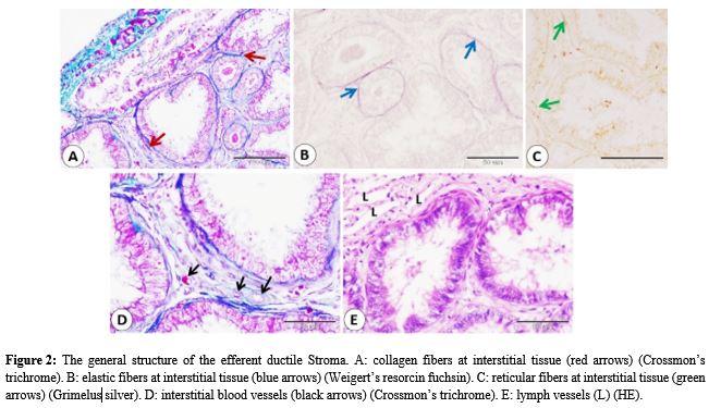
This research examines the structural features of the efferent ductules (EDs) in healthy doves (Spilopelia senegalensis) collected from local hunters in Assiut, Egypt. After slaughter, the efferent ductules were promptly dissected and preserved in Bouin's fluid. Utilizing a combination of histological, histochemical, and immunohistochemical techniques, we explored the architecture of EDs, focusing on the stroma and parenchyma. The lining epithelium was made up of a simple columnar to the pseudostratified columnar epithelium, ciliated, secretory, and basal cells. However, this study is the first to identify telocytes within the peritubular region of the dove’s efferent ductules, highlighting their potential role in tissue communication and regeneration. Additionally, androgen receptor expression was detected in the epithelial cells and spermatozoa within the lumen, suggesting a significant role in the male reproductive system.
Full text article
References
Abate-Shen, C., & Shen, M. M. (2000). Molecular genetics of prostate cancer. Genes & Development, 14(19), 2410–2434. DOI: 10.1101/gad.819500
Abd-Elmaksoud, A., Sayed-Ahmed, A., Mohamed, S. E., Mohamed, K., & Marei, H. (2009). Morphological and glycohistochemical studies on the epididymal region of the Sudani duck (Cairina moschata). Research in Veterinary Science, 86(1), 7–17. DOI: 10.1016/j.rvsc.2008.04.007.
Aire, T. (1980). The ductuli efferentes of the epididymal region of birds. Journal of Anatomy, 130(Pt 4), 707. PMCID: PMC1233197.
Aire, T. A. (2002). Morphological changes in the efferent ducts during the main phases of the reproductive cycle of birds. Journal of Morphology, 253(1), 64–75. DOI: 10.1002/jmor.1113
Aire, T., Ayeni, J., & Olowo-Okorun, M. (1979). The structure of the excurrent ducts of the testis of the guinea fowl (Numida meleagris). Journal of Anatomy, 129(3), 633. PMC1233029
Aire, T., & Soley, J. (2000). The surface features of the epithelial lining of the ducts of the epididymis of the ostrich (Struthio camelus). Anatomia, Histologia, Embryologia, 29(2), 119–126. DOI: https://doi.org/10.1046/j.1439-0264.2000.00247.x
Andraszek, K., & Smalec, E. (2011). The use of silver nitrate for the identification of spermatozoon structure in selected mammals. Canadian Journal of Animal Science, 91(2), 239–246. https://doi.org/10.4141/cjas10052
Bakst, M. (1980). Luminal topography of the male chicken and turkey excurrent duct system. Scanning Electron Microscopy, 3, 4194 - 4125. PMID: 7414286
Bancroft J. D., Layton, C., Suvarna, S. K. (2013). Bancroft's theory and practice of histological techniques. 7th ed. Amsterdam: Elsevier. Hardback ISBN: 9780702068645
Cretoiu, S. M., & Popescu, L. M. (2014). Telocytes revisited. Biomolecular Concepts, 5(5), 353–369. DOI: 10.1515/bmc-2014-0029
Earlé, R., & Dean, W. (1981). Features of spermatogenesis in the laughing dove Streptopelia senegalensis. African Zoology, 16(2), 109–112. https://www.ajol.info/index.php/az/article/view/152255
Gorgees, N. S., Habbib, O. A. A., & Hussein, R. A. (2013). The protective role of certain antioxidants (vitamins C, E and Omega-3) against aluminum chloride induced histological changes in the liver and kidney of female albino rats (Rattus Rattus Norvegicus). Science Journal of University of Zakho, 1(2), 591–603. https://sjuoz.uoz.edu.krd/index.php/sjuoz/article/view/276
Hess, R. A., Thurston, R., & Biellier, H. (1976). Morphology of the epididymal region and ductus deferens of the turkey (Meleagris gallopavo). Journal of Anatomy, 122(Pt 2), 241.
Hsu SM, Raine L, Fanger H. Use of avidin-biotin-peroxidase complex (ABC) in immunoperoxidase techniques: a comparison between ABC and unlabeled antibody (PAP) procedures. J Histochem Cytochem. 1981; 29:577–580. DOI: 10.1177/29.4.6166661
Ibrahim, M. I., Zakariah, M., Molele, R. A., Mahdy, M. A., Williams, J. H., & Botha, C. J. (2022). Ontogeny of the testicular excurrent duct system of male Japanese quail (Coturnix japonica): A histological, ultrastructural, and histometric study. Microscopy Research and Technique, 85(3), 1160–1170. DOI: 10.1002/jemt.23984
Ilio, K. Y., & Hess, R. (1994). Structure and function of the ductuli efferentes. Microsc Res Tech, 29, 432–467. DOI: 10.1002/jemt.1070290604
Madkour, F. A., & Mohamed, A. A. (2019). Comparative anatomical studies on the glandular stomach of the rock pigeon (Columba livia targia) and the Egyptian laughing dove (Streptopelia senegalensis aegyptiaca). Anatomia, Histologia, Embryologia, 48(1), 53–63.DOI: https://doi.org/10.1111/ahe.12411
Mustafa, F. E. Z. A., & El-Desoky, S. M. (2020). Architecture and cellular composition of the spleen in the Japanese Quail (Coturnix japonica). Microscopy and Microanalysis, 26(3), 589-598.
Mustafa, F. E. Z. A., & Elhanbaly, R. (2020). Distribution of estrogen receptor in the rabbit cervix during pregnancy with special reference to stromal elements: an immunohistochemical study. Scientific Reports, 10(1), 13655.
Mustafa, F. E.-Z. A., & Elhanbaly, R. (2021). Histological, histochemical, immunohistochemical and ultrastructural characterization of the testes of the dove. Zygote, 29(1), 33–41. DOI: https://doi.org/10.1017/S0967199420000477
Pawlicki, P., Yudakok-Dikmen, B., Tworzydlo, W., & Kotula-Balak, M. (2024). Toward understanding the role of the interstitial tissue architects: Possible functions of telocytes in the male gonad. Theriogenology. DOI: 10.1016/j.theriogenology.2024.01.013
Stefanini, M. A., Orsi, A. M., Gregório, E. A., Viotto, M. J. S., & Baraldi‐Artoni, S. M. (1999). Morphologic study of the efferent ductules of the pigeon (Columba livia). Journal of Morphology, 242(3), 247–255. DOI: 10.1002/(SICI)1097-4687(199912)242:3<247::AID-JMOR4>3.0.CO;2-G
Soliman, S. A., Abd-Elhafeez, H. H., Abou-Elhamd, A. S., Kamel, B. M., Abdellah, N., & Mustafa, F. E. Z. A. (2023). Role of uterine telocytes during pregnancy. Microscopy and Microanalysis, 29(1), 283-302.
Tingari, M. (1971). On the structure of the epididymal region and ductus deferens of the domestic fowl (Gallus domesticus). Journal of Anatomy, 109(Pt 3), 423. PMID: 4949288
Welsh, M., Moffat, L., Belling, K., De Franca, L., Segatelli, T., Saunders, P., Sharpe, R., & Smith, L. (2012). Androgen receptor signalling in peritubular myoid cells is essential for normal differentiation and function of adult Leydig cells. International Journal of Andrology, 35(1), 25–40. DOI: 10.1111/j.1365-2605.2011.01150.x
Yakirevich, E., Yanai, O., Sova, Y., Sabo, E., Stein, A., Hiss, J., & Resnick, M. B. (2002). Cytotoxic phenotype of intra-epithelial lymphocytes in normal and cryptorchid human testicular excurrent ducts. Human Reproduction, 17(2), 275–283. https://doi.org/10.1093/humrep/17.2.275. DOI: 10.1093/humrep/17.2.275.
Zhao, H., Yu, C., He, C., Mei, C., Liao, A., & Huang, D. (2020). The immune characteristics of the epididymis and the immune pathway of the epididymitis caused by different pathogens. Frontiers in Immunology, 11, 2115. DOI: 10.3389/fimmu.2020.02115
Zhu, L.-J., Hardy, M. P., Inigo, I. V., Huhtaniemi, I., Bardin, C. W., & Moo-Young, A. J. (2000). Effects of androgen on androgen receptor expression in rat testicular and epididymal cells: A quantitative immunohistochemical study. Biology of Reproduction, 63(2), 368–376. DOI: 10.1095/biolreprod63.2.368
Authors
Copyright (c) 2025 Fatma El-Zahraa Ahmed Mustafa, Basim. S. A. Al Sulivany

This work is licensed under a Creative Commons Attribution 4.0 International License.
Authors who publish with this journal agree to the following terms:
- Authors retain copyright and grant the journal right of first publication with the work simultaneously licensed under a Creative Commons Attribution License [CC BY-NC-SA 4.0] that allows others to share the work with an acknowledgment of the work's authorship and initial publication in this journal.
- Authors are able to enter into separate, additional contractual arrangements for the non-exclusive distribution of the journal's published version of the work, with an acknowledgment of its initial publication in this journal.
- Authors are permitted and encouraged to post their work online.

